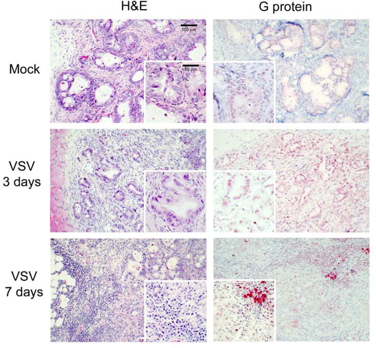
Figure 9.

Histological and immunohistochemical analysis of Panc 03.27 xenografts. Mock-infected tumors with injected with culture medium. M51R-VSV treated tumors received a single intratumoral injection (1×10⁸ pfu). Tumors were harvested three or seven days after treatment as indicated. Representative sections stained with hematoxylin and eosin (H&E) and immunohistochemical staining for VSV surface glycoprotein (G-protein) are shown from mock-treated and M51R-VSV.
