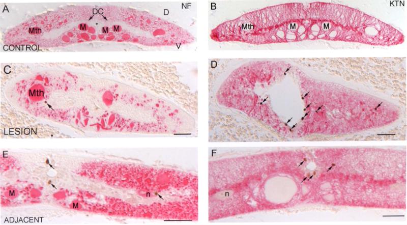
Fig. 2. BrdU label in the spinal cord is found in small cells, some labeling for glial keratin (KTN) but none for neurofilaments (NFs).
A and B, control lamprey spinal cord stained with anti-lamprey-NF (A) and anti-glial-keratin (B) antibodies. * = central canal; D = dorsal; V = ventral; Mth = Mauthner axon; M = Müller axon; DC = dorsal cell. C and D, lamprey spinal cord at 2 weeks post-TX and injected with BrdU 4 hours before sacrifice, stained with the anti-lamprey-NF antibody LCM3 (C) and the anti-lamprey-glial keratin antibody LCM29 (D). E and F, spinal cord 5 mm rostral (“adjacent”) to a TX, and processed as in C and D, stained with the same anti-lamprey-NF (E) and anti-lamprey-glial keratin (F) antibodies. Arrows point to BrdU-positive cells; scale bar = 50 μm. At this magnification, most glial cells are too small to definitively identify their nuclei with specific keratin-containing processes.

