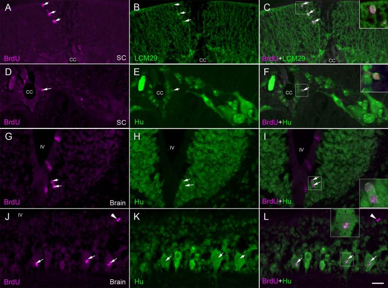
Fig. 8. Gliogenesis and neurogenesis after spinal cord transection.
At 2 weeks post-TX, animals were injected with BrdU and sacrificed 5 weeks later, in order to allow enough time for any post-mitotic cells to differentiate. Sections of the spinal cord and rhombencephalon were double stained for BrdU (A, D, G, J) and either keratin (B) or anti-Hu antibody (E, H, K). A, spinal cord at the TX site imaged for BrdU. B, same section imaged for keratin. C, superimposed. Note the co-labeling in glial cells (arrows) in the dorsal column. D, spinal cord imaged for BrdU. E, same section imaged for Hu. F, superimposed. Note the co-labeling in a cell (arrow; enlarged in inset) immediately subjacent to the cell layer lining the central canal (cc). Note the absence of co-labeling in the larger neurons away from the ependymal zone. G, Brainstem imaged for BrdU. H, same section imaged for Hu. I, superimposed. Note the co-labeling in ependymal cells (arrows; enlarged in inset). Label was also seen in cells of the subventricular zone (not shown here). J, Brain imaged for BrdU. K, same section imaged for Hu. L, superimposed. In a few larger neurons of the peripheral region, there is some BrdU labeling, but this does not occupy the entire nucleus (margins of the nucleus are indicated by arrows in the inset enlargement). It probably represents DNA repair (white arrows). Compare this pattern to that of the cells shown in the insets of C, F and I. White arrowheads point to a pair of BrdU-positive Hu-negative cells. Cc = central canal, SC = spinal cord, IV = fourth ventricle, scale bar = 20 μm.

