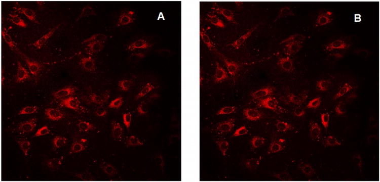
Figure 3.

K:Molv 3T3 NIH cells plated onto cover slips were incubated with CGlc4 for 24 hours, washed three times with PBS buffer, fixed with 4% paraformaldehyde, again washed three times with PBS, and mounted in Dako fluorescence mounting medium. Confocal images were taken using excitation wavelength 552 nm and emission filter was 578-700 nm. A: image after first scan, and B: image of the same sample after 25 scans. The images are as collected.
