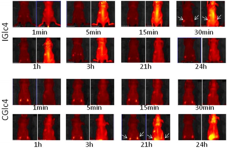
Figure 8.
IGlc4 and CGlc4 up-take, distribution, clearance. Four-week-old, male Balb/c athymic nude mice (Nu/Nu) were obtained from Harlan Sprague Dawley, Inc. (Indianapolis, IN). Mice were housed in temperature-controlled rooms (74 ± 2°F) with a 12-hour alternating light-dark cycle. Approximately 3×10⁶ GFP-JHU-22 cells in log phase were injected subcutaneously into the mice. The up-take experiments were done after seven days after GFP-JHU-22 cell inoculation. The mice received tail-vein injections with a total volume of 100 μLof 5mg/mLIGlc4 or CGlc4.The fluorescence images were taken at the times indicated after injection. Each individual group has two images where the emission due to GFP is on the left of each time interval, and monitored using a 465-520 band pass filter. A 605 -660 nm band pass filter was used to image the tumors with CGlc4 and IGlc4.The arrows indicated the location of tumor xenografts. A Xenogren IVIS instrument (Caliper Life Sciences, Hopkinton, MA) was used to collect these images.

