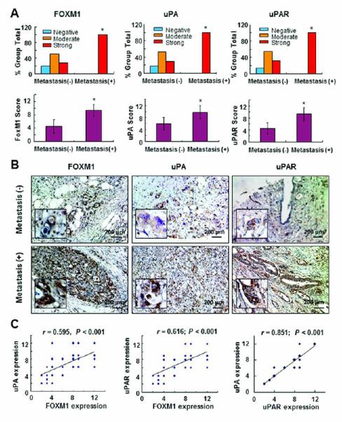
Figure 1.

Expression of FOXM1, uPAR, and uPA and its correlation with metastasis in pancreatic tumors. Staining of 70 primary pancreatic tumor specimens and 10 normal pancreatic tissue specimens for FOXM1, uPAR, and uPA protein was performed using a TMA. A, FOXM1, uPAR, and uPA expression was much higher in primary pancreatic tumor specimens obtained from patients with lymph node and/or distant metastasis (Metastasis [+]) than in those obtained from patients without metastasis (Metastasis [-]). *P < 0.05. B, representative photos of uPA, uPAR, and FOXM1 expression in primary pancreatic tumor specimens obtained from patients with or without metastasis at a magnification of ×200 (inserts, ×400). C, positive correlation of FOXM1 expression levels with uPA and uPAR expression levels in pancreatic tumor specimens (Pearson correlation test). Note: some of the dots on the graphs represent more than one specimen (overlapping scores).
