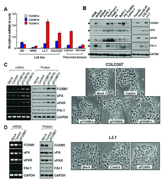
Figure 2.

Expression of FOXM1a, FOXM1b, and FOXM1c and its influence on EMT in pancreatic cancer cells. A, real-time PCR analysis using specific primers for FOXM1a, FOXM1b, and FOXM1c with pancreatic cancer cell lines and tumor specimens. FOXM1c was the predominant isoform expressed in HPDE cells and all pancreatic cancer cell lines, whereas FOXM1b was the predominant isoform expressed in HEK293 cells. The expression of FOXM1c in the highly metastatic L3.7 cells was much higher than that in the poorly metastatic COLO357 cells. Also, the expression of FOXM1c in pancreatic tumor specimens was much higher than that in adjacent normal pancreatic tissue specimens. B, Western blot analysis of FOXM1, uPA, uPAR, and PAI-1 protein expression in the pancreatic cancer cell lines and HPDE cells. C, COLO357 cells were transfected with pcDNA3.1-FOXM1a (pFOXM1a), -FOXM1b (pFOXM1b), or -FOXM1c (pFOXM1c) or with pcDNA3.1 alone and incubated for 48 hours. Forced expression of FOXM1b or FOXM1c in COLO357 cells greatly increased FOXM1, uPA, and uPAR mRNA and protein expression (left). Phase-contrast photomicrographs demonstrated that COLO357 cells underwent morphological changes typical of EMT after FOXM1b or FOXM1c overexpression (right). D, L3.7 cells were transfected with siFOXM1 or control siRNA and incubated for 48 hours. Forced siFOXM1 expression in these cells markedly inhibited FOXM1, uPA, and uPAR mRNA and protein expression (left). Phase-contrast photomicrographs demonstrated that L3.7 cells underwent morphological changes typical of mesenchymal-to-epithelial transition after knockdown of FOXM1 expression (right).
