
Fig. 10.
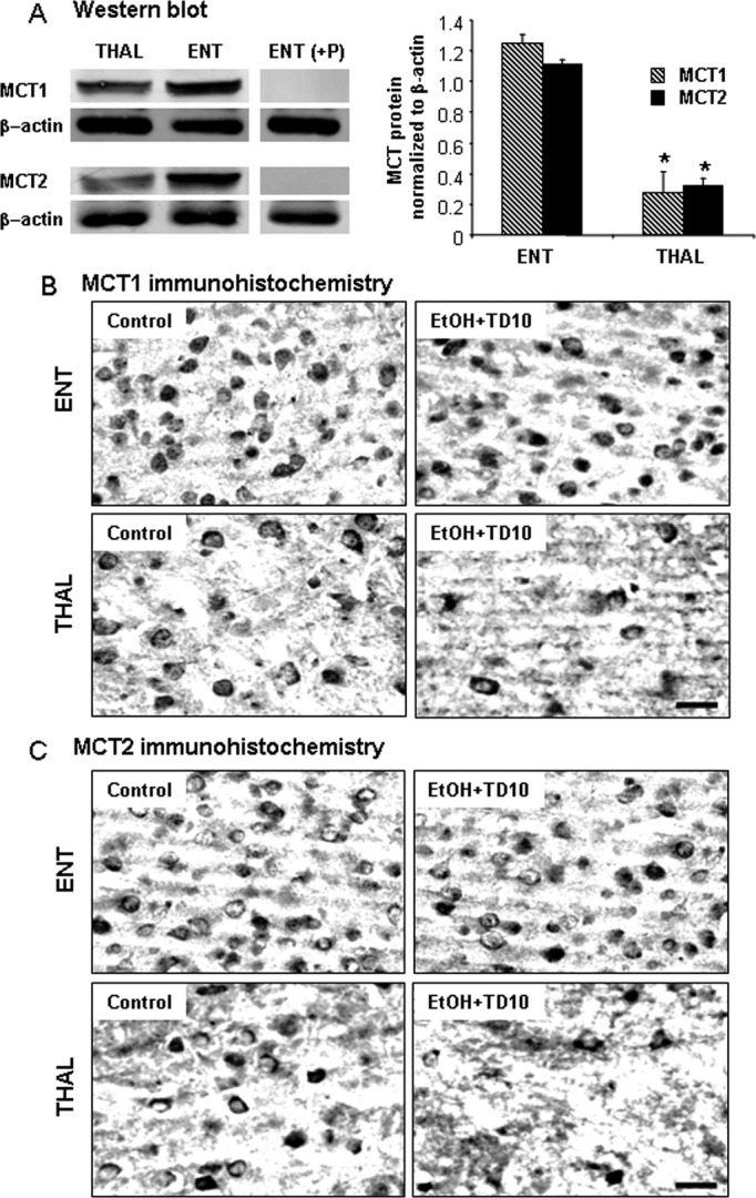
Monocarboxylic acid transporters (MCT1) and MCT2 expressions in Western blot and immunohistochemistry. (A) Thalamus (THAL) and entorhinal cortex (ENT) regions were dissected, and Western blot was performed as described in Methods. ENT shows more MCT1 and MCT2 protein expressions than THAL in untreated C57BL/6 mice. (B) C57BL/6 mice were treated with vehicle and EtOH+TD10 as described in Materials and Methods. Animals were sacrificed 24 hours following the last dose of EtOH treatment (5 g/kg, i.g. daily for 10 days). Brain sections were immunostained with MCT1 and MCT2 antibodies. EtOH+TD10-treated THAL showed decreased MCT1 + IR (B) and MCT2 + IR (C) cells, compared with the corresponding ENT. *P < 0.05, compared with ENT region. Scale bar = 50 μm.