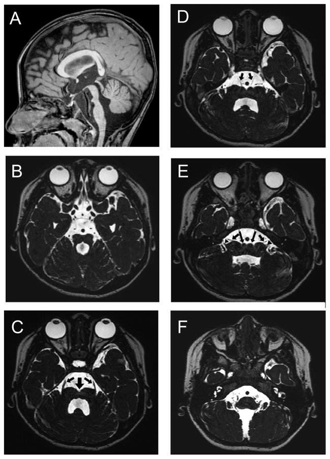
Figure 1.
Brainstem structures visualized on magnetic resonance imaging (MRI) of the patient at 2.5 years of age (A: T1-weighted sagittal section; B to F: constructive interference in steady-state sequences). A: The brainstem tegmentum appears hypoplastic at the level of the isthmus, and the protrusion of the ventral pons is attenuated. B: A molar tooth-like appearance is noted at the isthmus, composed by parallel orientation of superior cerebellar peduncles and an enlarged forth ventricle. B to E: A cleft (thick arrow) is seen at the midline along the entire length of the ventral pons. Small colliculi are noted on both sides of this cleft (long arrows in E). C: Entry of the trigeminal nerve roots (arrows) is dislocated to the most lateral end of the ventral pons. D: The abducens nerves (arrows) originate at the colliculli lateral to the midline cleft. E: The facial (arrows) and vestibulocochlear (arrowheads) nerves, which normally leave the brainstem in parallel and closely apposed to each other, originate separately.

