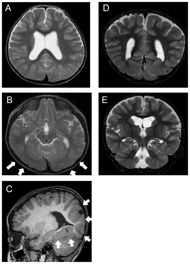
Figure 2.
Supratentorial structures on MRI (A, B, D and E: T2-weighted images; C: T1-weighted image). The cerebral cortex is thick in the posterior areas, including the bilateral temporal and occipital lobes (white arrows in B and C). The multi-convoluted cortical surface (C) and irregular gray-white matter boundary (B, C) are compatible with the findings in polymicrogyria. A myelinated layer is noted regionally within the thick cortex, which is suggestive of subcortical heterotopia (arrows in E). In D, hippocampal formation appears somewhat plump, but is not obviously dysplastic.

