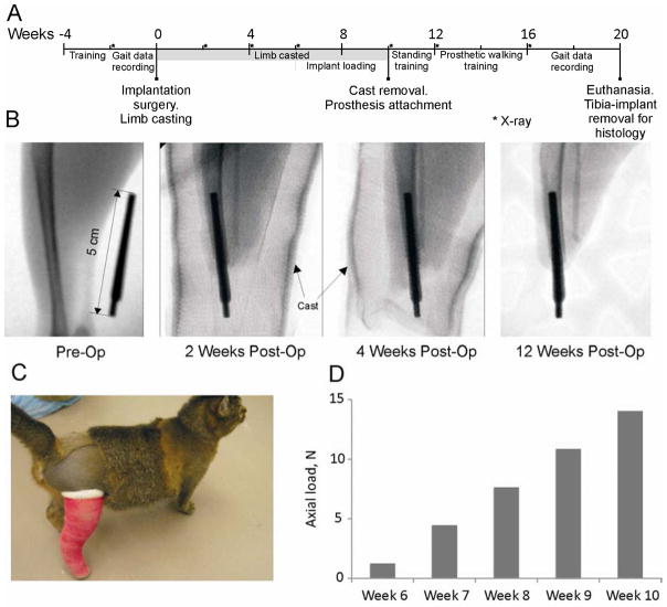
Figure 1.
Major stages of the study. A: Study timeline. Week 0 corresponds to the implantation surgery; the study ended with euthanasia and harvesting the residual limb with implant for histology (Week 21). B: X-ray images taken before surgery (Pre-Op), and 2, 4 and 12 weeks after implantation (Post-Op). C: Animal wearing a cast after pylon implantation to prevent premature loading of implant. D: Loads applied to implant using a handheld digital dynamometer from 6 to 10 weeks after implantation.

