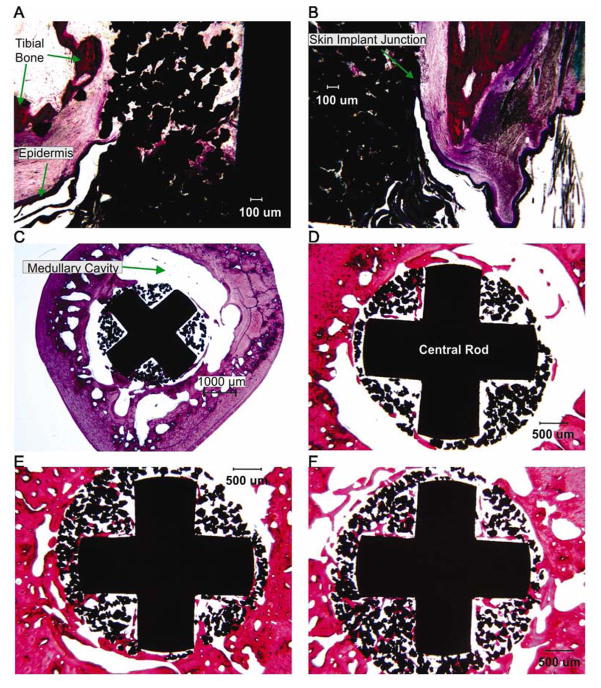
Figure 10.
Images of sections through implant, bone and skin from cat 2 (haemotoxylin and eosin stain). Locations of sections are shown in Fig. 9 A. A, B: Left and right views of the longitudinal section through the distal implant (40x magnification). C–F: Transverse cross sections through the bone and implant from proximal to distal sections, respectively (20x magnification).

