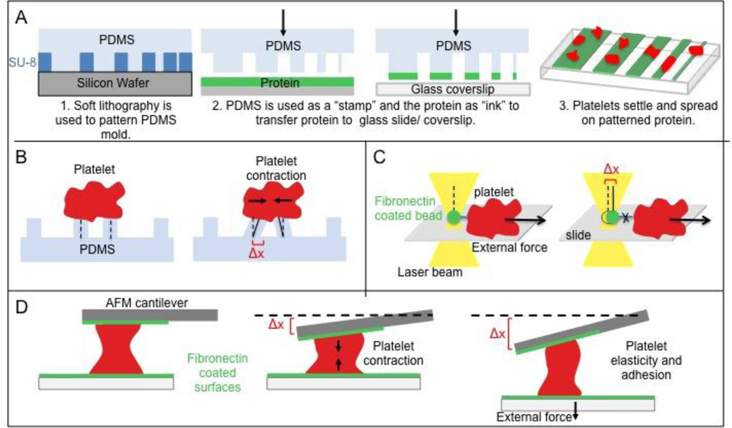
Figure 1. Tools of cell mechanics.
(A) Microstamping. Proteins are micropatterned to determine the dependency of a platelet on microenvironment geometry. (B) Platelet micropillar deflection. Platelets settle on PDMS pillars and pillar deflection (Δx) is used to compute platelet contractile force via Hooke’s Law, which states that force is directly proportional to Δx. (C) Optical tweezers or optical traps. A fibronectin-coated microbead held in a laser beam gradient is brought into contact with a platelet to allow for integrin binding. As the platelet is moved away from the bead, the bead deflection (Δx) at the point of bond rupture gives the strength of the αIIbβ3 – fibronectin bond. (D) Atomic force microscopy. Deflection of the AFM cantilever (Δx) gives platelet contractile force, or alternatively, platelet elasticity and adhesion if the bottom boundary is moved.

