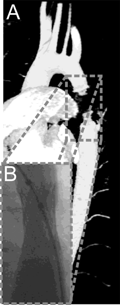
Figure 1.
Rendering of magnetic resonance imaging (MRI) data (A) and an angiographic image of the same patient obtained by fluoroscopy (B). CFD models were constructed using the MRI data with diameters of the stented region extracted from the fluoroscopy data to account for signal dropout created by artifacts due to the implanted Palmaz stent.

