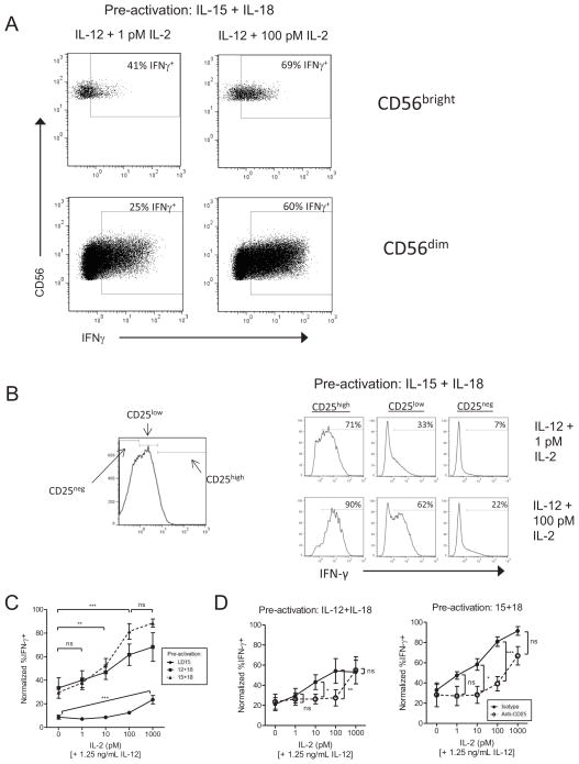
Figure 3. Induced CD25 on pre-activated NK cells results in a functional high affinity IL-2Rαβγ that signals for enhanced IFN-γ production.
Purified NK cells were activated for 16 hours with IL-12+IL-18 or IL-15+IL-18, washed extensively, and re-plated in cytokine-free media for 2 days to allow recovery to a resting state. IL-2 was then added at the indicated doses in combination with IL-12 (1.25 ng/mL). After 6 hours, IFN-γ was measured by intracellular flow cytometry. (A) Representative flow cytometric plots from one pre-activated donor depicting dose-dependent IFN-γ production co-stimulated by picomolar IL-2. (B) Representative flow plots from IL-15+IL-18 pre-activated NK cells that were subgated based on CD25 expression and IFN-γ expression with stimulation by IL-12 + 1 or 100 pM IL-2. (C) Summary data of pre-activated CD56dim NK cell IFN-γ production with IL-12 combined with various doses of IL-2, shown as mean ± SEM normalized IFN-γ percentage (n=4 donors). (D) In a separate assay, cells were pre-treated for one hour with either an anti-CD25 blocking antibody or IgG1 isotype control, followed by stimulation with 1.25 ng/mL IL-12 and 10-fold dilutions of IL-2. In (B,C), data is shown with the maximum IFN-γ positive percentage in each donor (n=4) normalized to 100%, since absolute IFN-γ+ percentages between donors are variable (range of maximum %IFN-γ+:14–66% for IL-12+IL-18 pre-activated cells, 28–86% for IL-15+IL-18 pre-activated cells). Significance was calculated by ANOVA.

