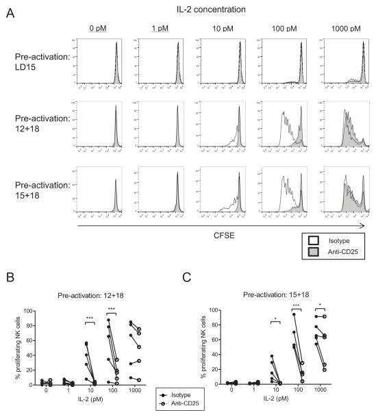
Figure 5. Low concentrations of IL-2 stimulate the proliferation of pre-activated CD56dim NK cells.
CFSE-labeled, flow sorted CD56dim NK cells were pre-activated with low dose IL-15, IL-12+IL18, or IL-15+IL-18 for 16 hours, washed extensively, and blocked with an isotype or anti-CD25 blocking antibody. Dilutions of IL-2 were then added, with subsequent media, cytokine, and blocking antibody additions occurring every other day. After 7 days, cells were harvested and analyzed for CFSE dilution by flow cytometry. Analysis was restricted to live cells by 7-AAD exclusion. (A) Data shown represents histograms from one donor. Both isotype-blocked (open histograms) and anti-CD25 blocked (shaded histograms) NK cell proliferation is shown. (B) Summary data depicting the variability of proliferation between CD56dim NK cells from different donors, and the dependence of picomolar IL-2 mediated proliferation on CD25 / IL-2Rαβγ. Significance was assessed by two-way ANOVA.

