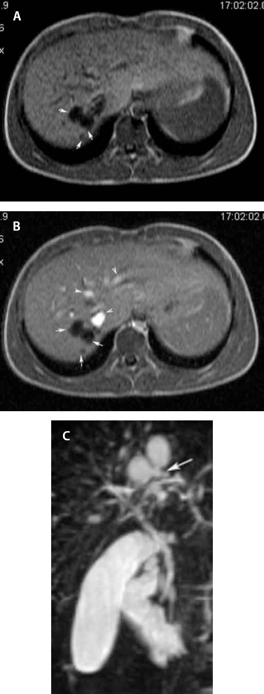
Figure 1.

a Plain T1-weighted axial scan demonstrates, in segment VII of the liver, three well defined round lesions of low signal intensity (arrows), b Contrast-enhanced T1-weighted axial scan shows no enhancement of the lesions (arrows); the enhancing structures represent vascular branches (arrowheads), c MRCP depicts the cysts communicating with the biliary tree (arrow).
