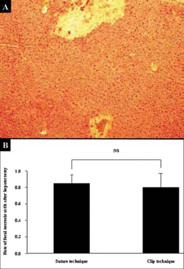
Figure 9.

Focal and patchy necrosis after massive hepatectomy. A. Focal and patchy necrosis was usually observed after massive hepatectomy. Findings by H-E staining at 100× magnification are shown. B. The rates of appearance of focal and/or patchy necrosis in hepatectomy were calculated from ten cases in each technique. Experiments were repeated three times. Histopathological analyses were performed at 6 h after 80% hepatectomy. There was no difference in the rate of appearance of focal and/or patchy necrosis between the suture technique and the clip technique (p = 0.6202).
