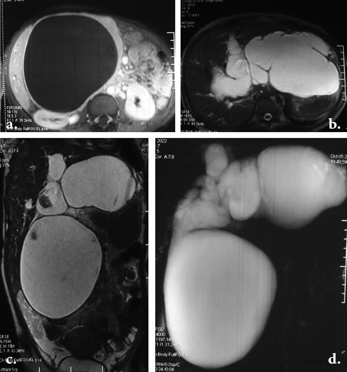
Figure 1.

a. Post-gadolinium axial T1W FSPGR image shows large cystic mass at the porta hepatis compressing the right kidney and other adjacent structures. b. Axial T2-weighted image above the level of confluence shows dilated left and right intrahepatic ducts with massive cystic dilatation of extrahepatic segment of the left hepatic duct extending to the left hypochondrium. The peripheral portions of intrahepatic ducts are not involved. c. Coronal T2-weighted image showing a huge cystic dilatation of the common bile duct extending to subhepatic area and another cyst in the epigastrium contiguous with the left hepatic duct. d. Projective MR image showing the bizarre cystic dilatation of the entire biliary tree.
