Figure 1.

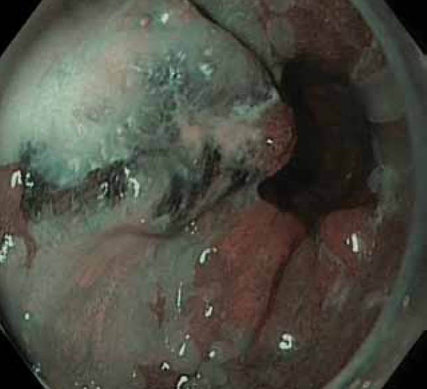
(B) Nodule in Barrett’s esophagus in Figure 1A seen with narrow band imaging: some overlying squamous mucosa clearly visible as well

(B) Nodule in Barrett’s esophagus in Figure 1A seen with narrow band imaging: some overlying squamous mucosa clearly visible as well