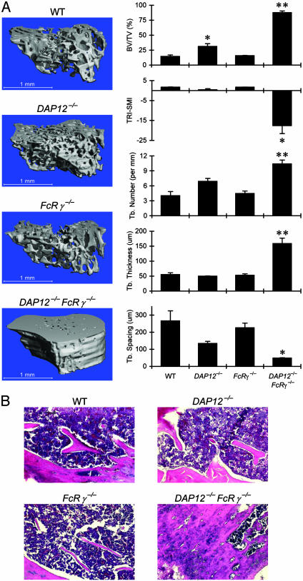
Fig. 1.
Osteopetrosis in mice lacking DAP12 and FcRγ.(A) Three-dimensional reconstitution of micro-CT scans of proximal tibia and 3D trabecular (Tb.) quantitative parameters (mean ± SEM) of bone structure. Significant differences from wild-type are shown (*, P < 0.05; **, P < 0.001). (B) Hematoxylineosin staining of decalcified sections of the primary spongiosa of proximal tibias. BV/TV, relative bone volume. TRI-SMI, 3D reconstruction image SMI.

