Figure 1.

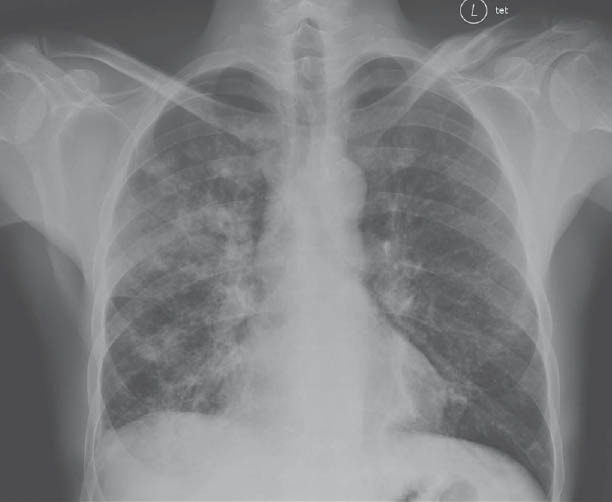
Initial chest radiograph. Increasing areas of confluent airspace disease involving the right middle and lower lobes and stable patchy airspace opacities involving both lungs with stable reactive lymphadenopathy

Initial chest radiograph. Increasing areas of confluent airspace disease involving the right middle and lower lobes and stable patchy airspace opacities involving both lungs with stable reactive lymphadenopathy