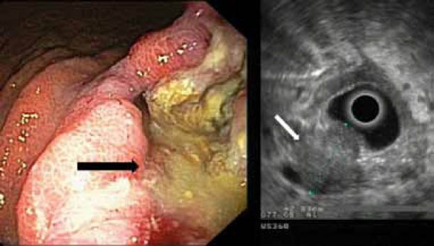
Figure 3.

A case of gastric cancer. Left: endoscopic image. Note the tumor that appears as a deep ulcer (black arrow). Right: EUS image of the same tumor (white arrow). Note the echo-poor, inhomogeneous appearance of the tumor, which spreads throughout the whole wall and its irregular outer margin (stage T3)
