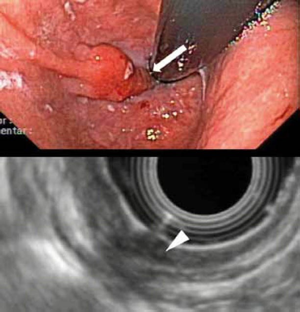
Figure 5.

EUS-staging of a T1 cancer in the gastroesophageal junction: Up: Endoscopic image. Note the tumor in retroflex imaging (arrow). Down: EUS-imaging of the tumor. Note the involvement of the 3 inner layers (T1sm), as well as the artefacts which make interpretation of the wall penetration by the tumor difficult
