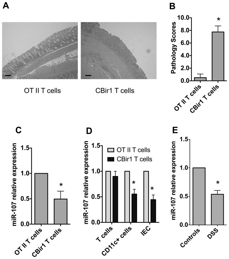
FIGURE 1. Decreased miR-107 expression in the inflamed intestine of mice with colitis.
1×10⁶ CD4+ T cells from CBir1 Tg mice or OT II mice were transferred into groups of 4 RAG−/− mice. Six weeks after transfer, the mice were sacrificed and the severity of intestinal inflammation was assessed by histological analysis and miR-107 expression was measured by real-time PCR. (A) Colonic histopathology of the recipient mice after HE staining is shown (bars, 100 μm). (B) Pathological scores are analyzed as previously described [22]. Data are shown as mean ± SD of eight mice pooled from two experiments *p<0.05, unpaired, two tailed Student’s t test. (C) RNA was isolated from the intestines of the recipient mice, and miR-107 expression in individual mice was determined by real-time PCR. Gapdh expression was used as a control. The miR-107 expression of RAG−/− mice receiving OT II T cells was arbitrarily set to 1.0, and used as the basis for calculating the relative changes in miR-107 expression. Data are shown as mean ± SD of four mice from one of two experiments performed. *p<0.05, unpaired, two tailed Student’s t test. (D) IEC, CD11C+ myeloid cells and T cells were isolated from the intestines of recipient mice and miR-107 expression was analyzed by real-time PCR. Gapdh was used as a house-keeping gene control. Data are shown as mean ± SD of four samples from one of two experiments performed. *p<0.05 compared with RAG−/− mice receiving OT II T cells, unpaired, two tailed Student’s t test. (E). Four C57BL/6 mice were fed with 2% DSS for 7 days and then water for 3 days. Control mice were fed with water only. miR-107 expression in the intestines was determined 10 days later by real-time PCR. Data are shown as mean ± SD of four samples from one of two experiments performed. *p<0.05, compared with control C57BL/6 mice, unpaired, two tailed Student’s t test.

