Fig. 2.
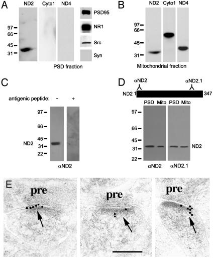
ND2 is present at the PSD. (A) Immunoblots of PSD proteins probed with anti-ND2, anti-cytochrome c oxidase I (Cyto1), anti-ND4, anti-PSD95, anti-NR1, anti-Src, and anti-synaptophysin. The PSD preparation contained PSD95, NR1, and Src, but lacked the presynaptic marker synaptophysin. (B) Immunoblots of mitochondrial proteins probed with anti-ND2, anti-Cyto1, and anti-ND4. Neither NR1 nor NR2A/B was detected in the mitochondrial fraction (not shown). (C) Immunoblots of PSD proteins showing the specificity of the N-terminal ND2 antibody by preadsorption with the antigenic peptide used to derive the antibody. (D) Immunoblots of PSD and mitochondrial proteins probed with two independent rabbit polyclonal antibodies directed against two disparate regions of ND2. The N-terminal ND2 antibody was used for all subsequent experiments shown. (E) Postembedding immunogold electron microscopy images of rat hippocampus CA1 synapses. ND2 immunoreactivity in PSDs is visualized by secondary antibody conjugated to 10-nm gold particles. pre, presynaptic. (Scale bar is 200 nm.)

