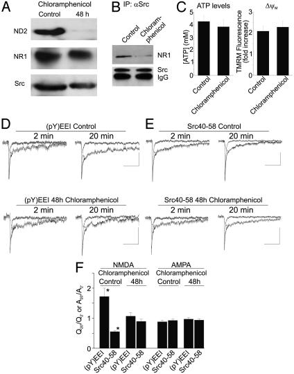
Fig. 5.
Blocking expression of ND2 prevents Src-dependent regulation of NMDA receptor activity. (A) Immunoblots of total soluble protein obtained from cultured rat hippocampal neurons treated with 50 μg/ml chloramphenicol (CAP) for 48 h probed with anti-ND2, anti-NR1, and anti-Src as indicated. (B) Immunoblot of coimmunoprecipitates obtained from cultured hippocampal neurons, either untreated or treated with CAP for 48 h, probed with anti-NR1 or anti-Src. (C) Summary histograms (Left) of ATP level or mitochondrial membrane potential (▵ψM), as assessed by TMRM fluorescence dequenching (Right), in cultured hippocampal neurons either untreated or treated with CAP for 48 h. (D) The up-regulation of NMDAR activity by the Src activator peptide EPQ(pY)EEIPIA, labeled (pY)EEI, is prevented in CAP-treated neurons. (E) The reduction of NMDAR activity by Src40–58 peptide is also prevented in neurons treated with CAP for 48 h. Composite traces are shown in black, the NMDAR component is shown in dark gray, and the AMPA receptor (AMPAR) component is shown in light gray. (Scale bars are 50 ms/10 pA.) (F) Summary histogram of electrophysiology data. NMDA component data were calculated as Q20′/Q2′, and AMPA component data were calculated as A20′/A2′. CAP treatment for 48 h prevents modulation of NMDAR function by the Src activator and Src40–58 peptides, whereas neither of these reagents affected the AMPAR component of mEPSCs. Asterisks indicate a significant difference, Student's t test, P < 0.05.

