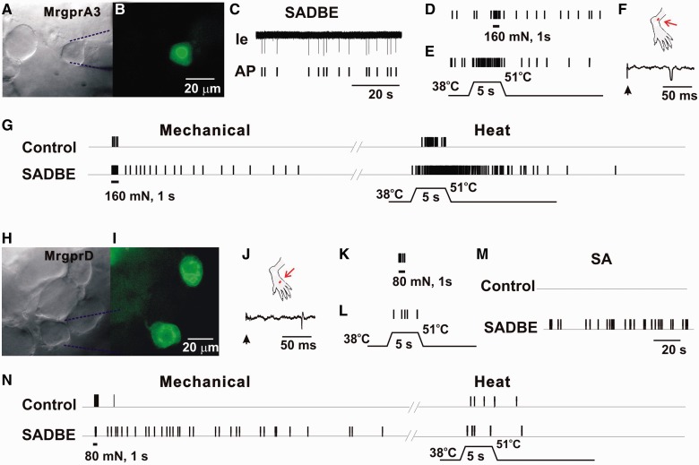
Figure 2.
CHS produced spontaneous activity and enhanced stimulus-evoked responses in MRGPRA3+ and MRGPRD+ neurons. (A) Bright-field image of a neuron under recording with the extracellular electrode (outlined with dashed blue lines). (B) Fluorescent microscopy revealed the expression of GFP (i.e. MRGPRA3) in this neuron. (C) Original extracellular recording trace (Ie) and action potential markers (AP) indicated the presence of abnormal spontaneous activity (SA) of this neuron, without any external stimulation. (D) Responses of this MRGPRA3-GFP+ neuron to 160 mN force through a 200 μm diameter probe (1 s). (E) Response to heat stimulation (38 to 51°C, 5 s), in the presence of ongoing spontaneous activity. (F) Location of cutaneous receptive field (red dot) of this neuron on the hairy skin of hind paw (the region challenged with SADBE), and conduction velocity (0.67 m/s, lower trace) obtained with electrical stimulation (arrow) to the peripheral receptive field. (G) Responses of the MRGPRA3+ neuron innervating the vehicle control or SADBE-challenged skin to mechanical stimulus of 160 mN force through a 200 μm probe for 1 s, and to heat stimuli (38 to 51°C, 5 s). The neuron innervating SADBE-challenged skin was initially silent but showed prolonged after-discharge following the 160 mN mechanical stimulation and heating. No stimulus-evoked after-discharges were observed in MRGPRA3 neurons from control animals. (H) Bright-field image of a MRGPRD+ neuron under recording with the extracellular electrode (outlined with dashed blue lines). (I) Fluorescent microscopy revealed the expression of GFP (or MRGPRD) in this neuron. (J) Location of cutaneous peripheral receptive field (red dot) of this neuron on the hairy skin of hind paw (the region challenged with SADBE), and conduction velocity (0.57 m/s, lower trace) obtained with electrical stimulation (arrow) to the peripheral receptive field. (K and L) Responses of MRGPRD-GFP+ neuron to 80 mN force through a 200 μm diameter probe (1 s), and to heat stimulation (38 to 51°C, 5 s). This neuron did not exhibit spontaneous activity. (M) Spontaneous activity was observed in a subset of MRGPRD+ neurons from CHS mice, but not from controls. (N) MRGPRD+ neurons from CHS mice exhibited prolonged after-discharges in response to noxious mechanical stimulus (80 mN, 1 s) but not heat (38 to 51°C, 5 s). No prolonged after-discharges were evoked by noxious mechanical and heat stimuli in MRGPRD+ neurons from control animals.

