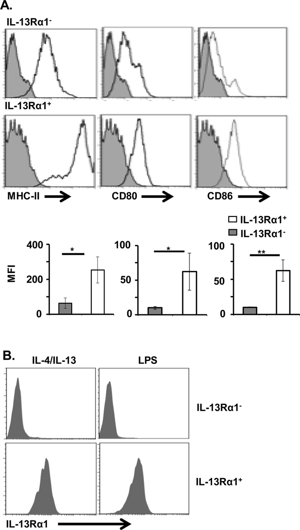
Figure 3.
IL-13Rα1 is stably expressed on macrophages that display high levels of MHC and costimulatory molecules. Purified splenic CD11b+F4/80+IL-13Rα1+ and CD11b+F4/80+IL-13Rα1− macrophages from IL-13Rα1+/+-GFP mice were stained for MHC II and costimulatory molecules and analyzed by flow cytometry. (A) Expression of MHC II, CD80 and CD86 on IL-13Rα1+ and IL-13Rα1− Macrophage populations. The upper panel shows representative flow cytometry data from 5 experiments, while the bottom panel shows mean ± SD of MFI data compiled from 5 experiments. *p < 0.05, ** p< 0.005 (unpaired two-tailed student t test). (B) IL-13Rα1+ and IL-13Rα1− Macrophage populations were stimulated with a mixture of IL-4 and IL-13 (30 ng/mL of each cytokine), or LPS (100 ng/mL) for 24 h and GFP (IL-13Rα1) expression was analyzed by flow cytometry.

