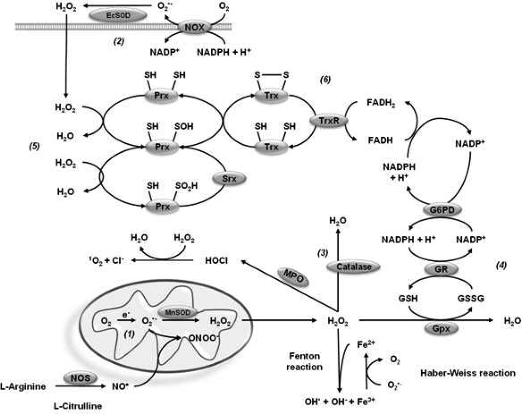
Figure 6. Catalytic antioxidant systems.
Cells have intrinsic antioxidant mechanisms to maintain a tight homeostatic control of ROS/RNS generated under physiological conditions, and also detoxify their excessive accumulation in pathological conditions. SODs dismutate O2•− to O2 and H2O2. (1) MnSOD is localized in the mitochondrial matrix, CuZnSOD in the IMS, peroxysomes, nucleous or cytosol, and EcSOD in the extracellular space (2). (3) Catalase catalyzes the decomposition of H2O2 to water and oxygen and is primarily localized in the peroxisomes. (4) Gpx are selenoproteins that reduce peroxides using GSH as substrate. Gpx isozymes encoded by different genes vary in their cellular location and substrate specificity. For example, Gpx1 preferably scavenges H2O2, whileGpx4 hydrolyses lipid hydroperoxides. GR, which reduces GSSG back to GSH, requires NADPH as electron donor reductant, and G6PD is indispensable for the regeneration of NADPH from NADP+ in then cytosol. (5) Prxs are ubiquitous thiol peroxidases that scavenge peroxides. Catalysis of H2O2 is mediated by the reaction of their active site cysteine residue with H2O2 with a rate comparabale to that of catalases. The catalytic activity of Prxs requires reducing equivalents provided by the Trx/TrxR system. (6) In addition, Trxs can directly bind to proteins modifying their function.

