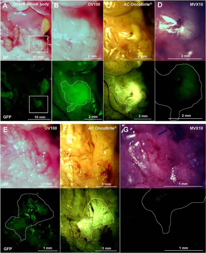
Fig. 4.
Imaging of PDOX tumor labeled with fluorescent anti-CA19-9 antibody. The primary PDOX tumor, labeled with fluorescent anti-CA19-9 antibody, was imaged with the OV100 at a magnification of 0.14× and 0.56× (A and B), the Dino-Lite at a magnification of 30× (C) and the MVX10 at a magnification of 1.6× (D). The MVX10 at a magnification of 3.2× did not detect any signals from the residual tumor (G). The OV100 at a magnification of 0.89× could not distinguish the residual tumor from background (E). The Dino-Lite at a magnification of 50× could clearly distinguish the residual tumor from background (F). Boxes in (A) indicate the view areas of (B), (C) and (D). The areas surrounded by white broken lines indicate the estimated residual tumor areas after BLS.

