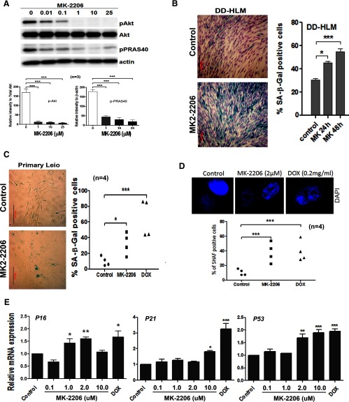
Figure 1.
Inhibition of AKT induces senescence. A, Western blot analysis of pAKT and its downstream effector (pPRAS40) in primary leiomyoma cells treated from left to right: DMSO (0), MK-2206 0.01, 0.1, 1, 10, 25 μM. Relative expression levels of pAKT and pPRAS40 were shown below. B, Senescence detected by SA-β-Gal in DD-HLM (right) treated with MK-2206 in 24 and 48 hours (left). C, Senescence analysis in primary leiomyomas cells (n = 4) in control, MK-2206 (2 μM), and DOX (0.2 μg/mL) treatment. D, SAHF observed by florescent microscope (upper panel) and dotplot analysis of SAHF (bottom panel) in primary leiomyoma cells (n = 4) treated with MK-2206 or DOX. E, Leiomyoma cell lines were treated with increasing doses of MK-2206 and expression analysis of senescence-associated genes P16, P21, and P53 was measured by real time RT-PCR. The data shown are means ± SEM from 3 independent experiments (n = 3). *, P < .05; **, P < .01; ***, P < .001.

