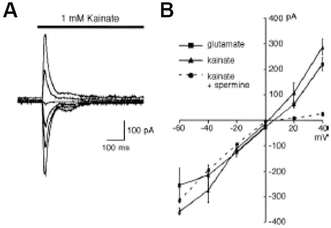
Figure 4. Ligand-gated currents in AVA neurons.

Currents activated by application of the indicated ionotropic glutamate receptor agonists. (A) kainate-gated membrane current recorded from AVA in vivo. Membrane potential was varied between -60 and +40 mV (in 20mV increments). (B) Peak current-voltage relationship for glutamate- and kainate-gated currents. As found in mammalian neurons, intracellular spermine blocks outward current. Reprinted with permission (Mellem et al., 2002).
