
Figure 5. Light-activated post-synaptic potentials in AVA neurons.
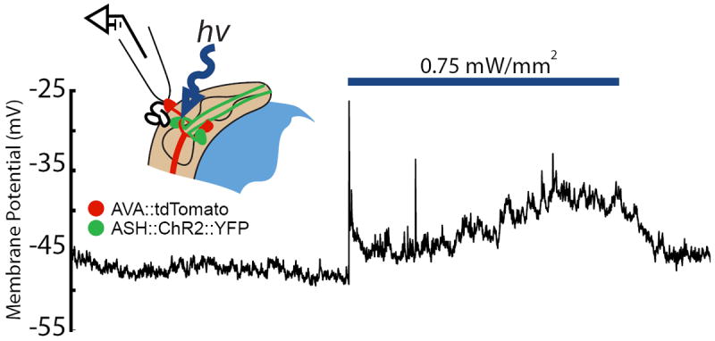
A light pulse (blue bar) activates the ASH neuron, which is pre-synaptic to AVA and expresses ChR2∷YFP, and induces a post-synaptic potential in the AVA neuron (labeled with tdTomato). The configuration for recording light-activated synaptic currents is illustrated in the inset. (T. Lindsay, S. Lockery, unpublished).