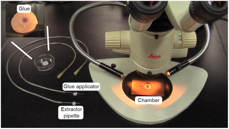
Figure 8. NMJ dissection apparatus.

The dissection microscope (total magnification 120X) is equipped with a black base and illuminated with gooseneck fiber optics for optimal visualization during dissection. The glue applicator and viscera extractor are similarly constructed and are mounted on dental wax between uses. Since the extractor becomes fluid filled during suction, where as the glue applicator must remain free of fluid for accurate glue application, the extractor is kept in the front position at all times to avoid confusion. The recording chamber, shown on the microscope base has a PDMS-coated coverslip inserted into the central chamber and held in place by a wax ring. The insert shows a close-up of the glue container, showing a drop of HistoacrylBlue glue, contained within a PCR tube lid, embedded in dental wax.
