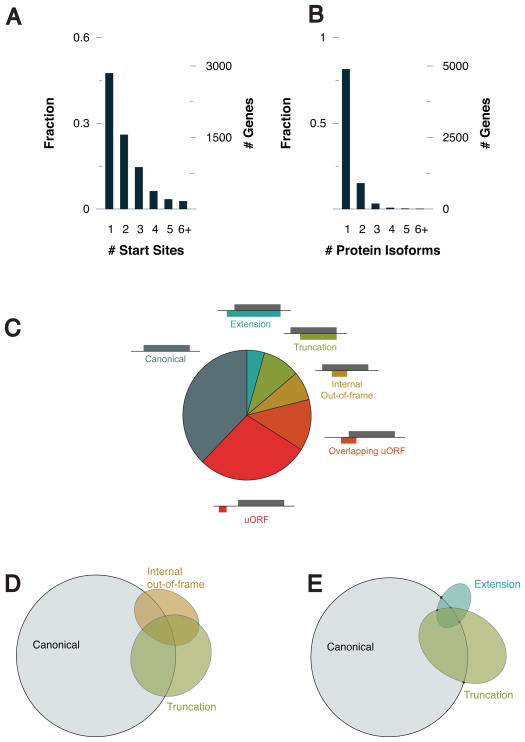
Figure 6. Ribosomal profiling of human monocytes identifies extension and truncation variants similar to miniMAVS.
(A) The fraction and number of genes that were detected to have one or more translational start site.
(B) The fraction and number of genes that have more than one translational start site resulting in either an extension or truncation.
(C) Classification of each of start site relative to the reading frame of the annotated CDS.
(D) Venn diagram showing the number of genes identified containing one or more canonical, truncation, or internal out-of-frame start site.
(E) Venn diagram showing the number of genes identified containing one or more canonical, truncation, or extension start site.

