
Figure 4.
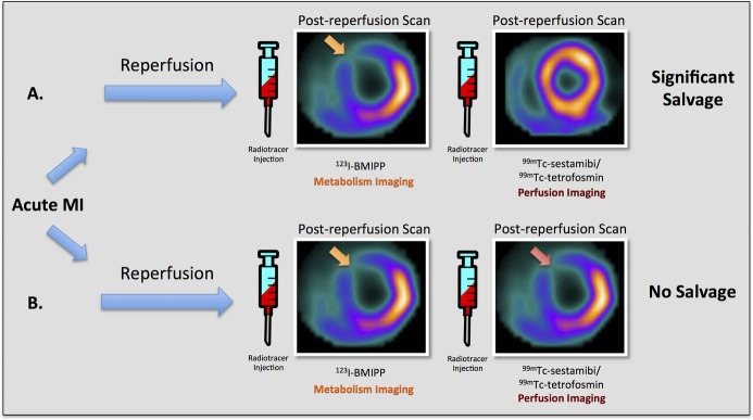
SPECT myocardial metabolism/perfusion imaging for the assessment of at‐risk myocardium. Diagram depicting combined fatty acid SPECT metabolic imaging with 123I‐BMIPP and technetium‐based SPECT perfusion imaging with 99mTc‐sestamibi/tetrofosmin for the assessment of myocardial salvage in 2 different hypothetical examples of acute ischemic myocardial injury in the left anterior descending (LAD) coronary artery territory. This technique requires injections of 2 separate radiotracers (123I‐BMIPP and 99mTc‐sestamibi/tetrofosmin) following reperfusion. A, Post‐reperfusion 123I‐BMIPP SPECT imaging reveals an area of decreased counts in the anteroseptum and anterior wall, whereas post‐reperfusion 99mTc‐sestamibi/tetrofosmin SPECT imaging shows normal myocardial perfusion. These findings suggest significant myocardial salvage in the left anterior descending coronary artery territory. B, Both post‐reperfusion 123I‐BMIPP SPECT metabolic imaging and post‐reperfusion 99mTc‐sestamibi/tetrofosmin perfusion SPECT imaging demonstrate decreased counts in the anteroseptum and anterior wall, suggesting no myocardial salvage in the left anterior descending coronary artery territory. 123I‐BMIPP indicates β‐methyl‐p‐[123I]‐iodophenyl‐pentadecanoic acid; 99mTc, technetium‐99m; MI, myocardial infarction; SPECT, single‐photon emission computed tomography.