
Figure 5.
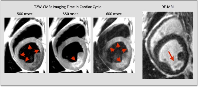
Technical issues with T2‐weighted CMR. Technical limitations with conventional dark‐blood T2W‐CMR. Images taken from a subject with myocardial infarction in the left circumflex coronary artery territory. Red arrowheads indicate areas of myocardium with high signal by T2W‐CMR. The red arrow indicates a subendocardial lateral wall infarct on corresponding DE‐CMR image at the same slice location. Note that the regional myocardial signal intensity varies significantly with small changes in the acquisition time of the cardiac cycle. (Adapted and reprinted, with permission, from reference no. 40.) DE‐CMR indicates delayed‐enhancement cardiac magnetic resonance; MRI, magnetic resonance imaging; T2W‐CMR, T2‐weighted cardiac magnetic resonance.