
Figure 6.
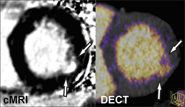
Cardiac magnetic resonance imaging (cMRI) and dual‐energy computed tomography (DECT) imaging of myocardial infarction. Iodine color map reconstructed from a DECT image demonstrates good agreement when compared with delayed‐enhancement cMRI of a subject with prior myocardial infarction of the lateral wall. Similar to cMRI, DECT offers the potential to assess myocardial infarction as well as myocardial salvage through the use of pre‐ and postcontrast techniques.