Figure 2.

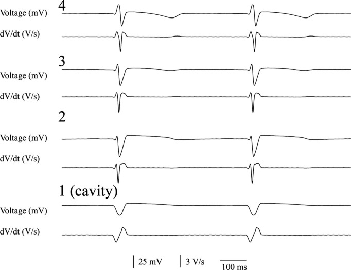
Voltage electrograms (top) and their first temporal derivative (bottom) from the 4 most distal electrodes from a plunge needle during sinus rhythm. The most distal electrode (1) was in the ventricular cavity.

Voltage electrograms (top) and their first temporal derivative (bottom) from the 4 most distal electrodes from a plunge needle during sinus rhythm. The most distal electrode (1) was in the ventricular cavity.