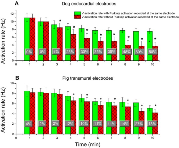
Figure 9.

Mean and SD of WM activation rates in recordings with and without Purkinje activations every minute during VF. A, is for the most endocardial electrodes of each plunge needle in dogs. B, for all plunge needle electrodes within the ventricular wall in pigs. Percent changes of activation rate at each minute between recordings with and without Purkinje activations during VF are superimposed on the bar graphs. An asterisk indicates P<0.05 for that minute of VF. VF indicates ventricular fibrillation; WM, working myocardium.
