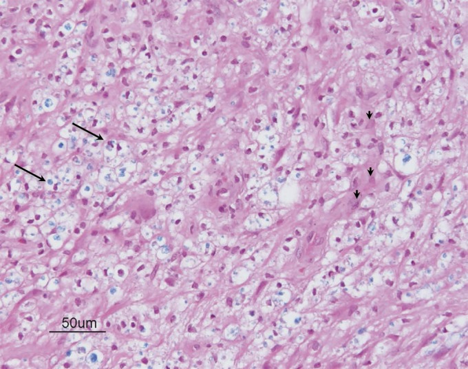
Figure 2. Spinal cord biopsy.

Hematoxylin & eosin/Luxol fast blue (LFB) stained section from the spinal cord biopsy demonstrates sheets of macrophages (arrows) containing LFB-positive debris and scattered reactive astrocytes (arrowheads) suggestive of an active demyelinating process (200×).
