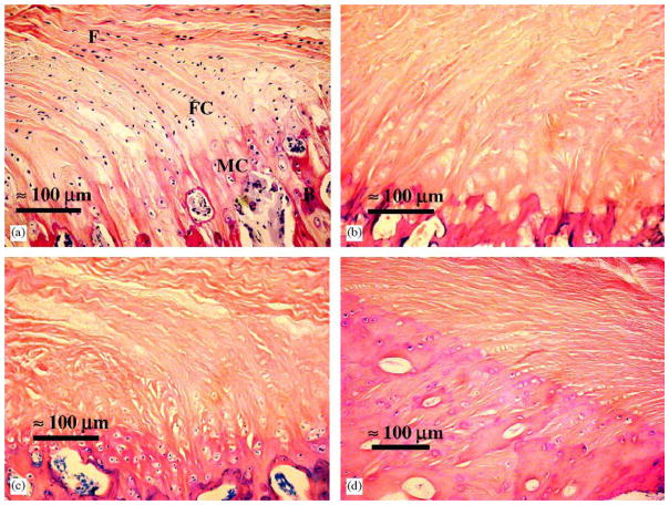
Figure 3.
Decellularized and untreated ACL and its tibial insertion site in porcine samples stained with hematoxylin and eosin at 20× magnification. (a) Untreated ACL with collagen fibers (F) passing through fibrocartilage (FC), mineralized cartilage (MC) and bone (B). (b) ACL treated with Triton–SDS. (c) ACL treated with Triton–Triton. (d) ACL treated with Triton–TnBP. Scale bars: 100 μm. Reprinted from Woods et al. 2005 with permission from Elsevier Publisher, Ltd.
Porcine tibial insertion of decellularized and untreated Anterior Cruciated Ligaments.

