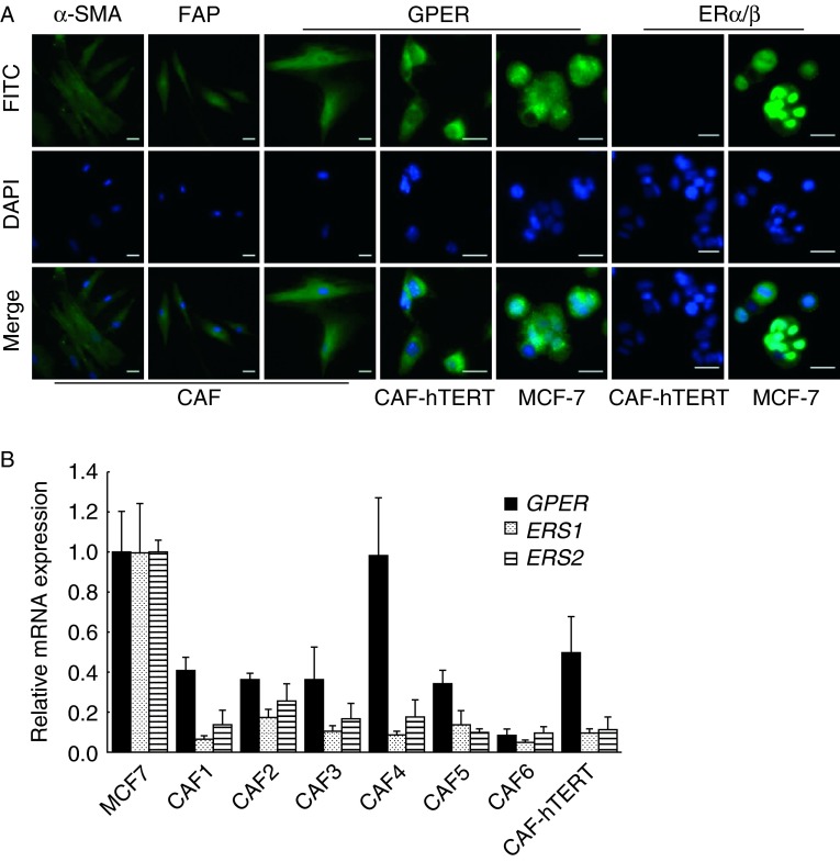
Figure 2.
GPER is expressed in primary and immortalized breast CAFs. (A) CAFs were identified by immunofluorescent staining with α-SMA and FAP. ERs were detected in primary breast CAFs, immortalized CAFs (CAF-hTERT), and MCF-7 cells as positive controls. Scale bars: 25 μm. (B) ER mRNA expression was evaluated by quantitative real-time RT-PCR in primary CAFs, CAF-hTERT cells, and MCF-7 cells. Gene expression was normalized to GAPDH, and results are shown as fold changes of mRNA levels compared with MCF-7 cells. The data are shown as means±s.d. for three independent experiments.

 This work is licensed under a