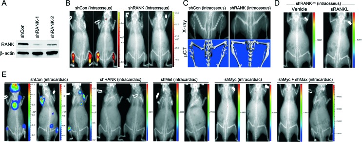
Figure 6.
Abrogation of RANK or its downstream target transcription factors, c-Myc/Max, or its effector/target, c-Met abolishes the metastatic potential of LNRANKL cells. (A) Partial RANK knockdown in LNRANKL cells was confirmed by western blot analysis. (B) Representative NIR fluorescent images showed that RANK-knocked-down LNRANKL cells (n=10) form no or very small tumors undetectable by NIR fluorescent imaging, but LNRANKL cells transduced with control shRNA (shCon) (n=10) form obvious intratibial tumors (average total radiant efficiency of NIR ((p/s)/(μW/cm2)): shCon (intraosseus), 4.86×10¹⁰ and 3.37×10¹⁰; shRANK (intraosseus), 2.63×10⁵ and 3.84×10⁵). (C) X-ray and μCT scans showed that LNRANKL cells induced osteolytic lesions in mouse tibia, unlike RANK-knocked-down LNRANKL cells where mouse tibia were intact with minimal detectable bone lesions. (D) Representative in vivo bioluminescent images further demonstrated that administration of recombinant sRANKL (50 μg/kg s.c. twice a week, 2 weeks after intraosseous inoculation of test cell line) fails to induce intratibial tumor formation of luciferase-tagged RANK-knocked down LNRANKL cells in mice (n=10) (total flux (photons/s): shRANK-1 (intraosseus) vehicle, 2.53×10³ and shRANK-1 (intraosseus)+sRANKL, 1.80×10⁴). (E) Representative in vivo bioluminescent images further demonstrated that luciferase-tagged LNRANKL shCon cells, but not luciferase-tagged LNRANKL cells with RANK (n=10), c-Met (n=10), c-Myc (n=8), or c-Myc/Max (n=8) knockdown, induce bone and soft tissue metastases after intracardiac injection (average total flux (photons/s): shCon (intracardiac), 4.60×10⁸, 9.71×10⁶, and 4.01×10⁷; shRANK (intracardiac), 4.27×10³ and 6.13×10³; shMet (intracardiac), 4.52×10³ and 2.25×10⁴; shMyc (intracardiac), 8.51×10³ and 1.83×10⁴; and shMyc+shMax (intracardiac), 8.65×10⁴ and 1.64×10⁵).

 This work is licensed under a