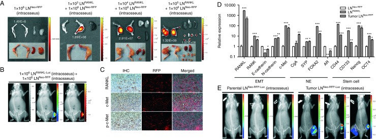
Figure 7.
Metastatic LNRANKL cells can transform neighboring non-metastatic LNNeo cells to undergo EMT, express neuroendocrine (NE) and stem cell biomarkers, and form tumors in mouse skeleton. (A) A small population of LNRANKL cells was capable of recruiting non-metastatic LNNeo-RFP cells, which were incapable of forming tumors by themselves, to participate in bone colonization in mice. One thousand, ten thousand, and one hundred thousand LNRANKL cells were co-inoculated with one million LNNeo-RFP cells in both tibia of nude mice. Tibial tumors and tumors disseminated from bone to soft tissues were harvested and subjected to fluorescent imaging to detect the red fluorescent signal from non-metastatic LNNeo-RFP cells using the Xenogen imaging system at an excitation of 535 nm and emission of 620 nm. Spleens were also harvested and used as negative controls. Total radiant efficiency ((p/s)/(μW/cm2)) of the RFP in tumors was labeled in white in the corresponding tumors. (B) Representative in vivo bioluminescent and red fluorescent images were also demonstrated with mice bearing intratibial inoculation of either luciferase-tagged LNRANKL cells followed by intracardiac inoculation of LNNeo-RFP cells to test the homing potential of RFP-tagged LNCaPNeo cells. (C) IHC and fluorescence images were obtained from chimeric tumors induced in mouse skeleton by inoculating 1000 LNRANKL cells plus 1×10⁶ LNNeo-RFP cells. Representative IHC and fluorescence images of the tumors were merged (200× magnification). Data show co-localization of RFP cells with RANKL, c-Met, and p-c-Met expression in prostate tumors from mouse skeleton. (D) LNNeo-RFP cells harvested from chimeric tumors (tumor LNNeo-RFP) acquired EMT, neuroendocrine, and stem cell properties demonstrated by relative expression of markers detected by qRT-PCR (*P<0.05; **P<0.01; and ***P<0.001). (E) Representative bioluminescent images demonstrated tibial tumor formation induced by luciferase-tagged tumor-derived LNNeo-RFP cells but not by luciferase-tagged parental LNNeo-RFP cells (total flux (photons/s): parental LNNeo-RFP-Luc, 5.77×10³; 2.09×10⁴; and 1.56×10⁴ and tumor LNNeo-RFP-Luc, 5.35×10⁶; 9.94×10⁵; and 8.52×10⁵).

 This work is licensed under a