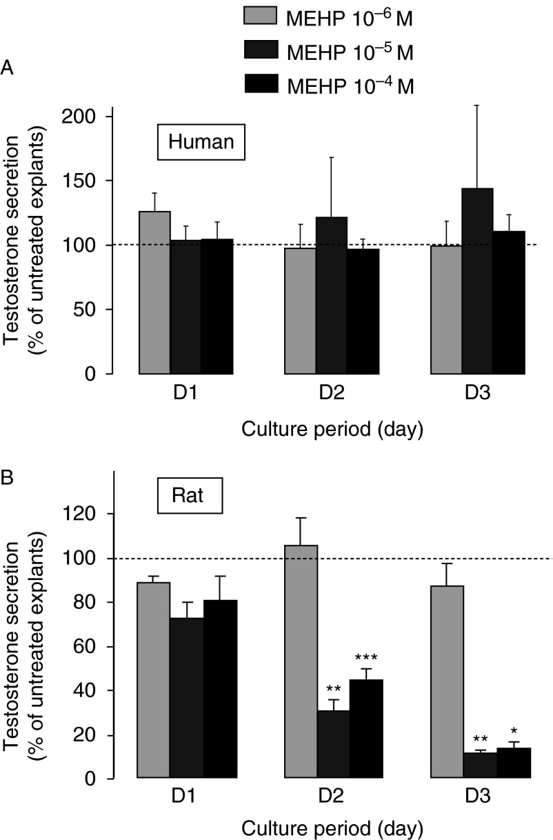
Figure 4.
Effect of MEHP on testosterone secretion by cultured rat and human fetal testis explants. Testes from 7 to 11 GW human fetuses (A) to 14.5-day-old rat fetuses (B) were cultured using the FeTA system, a method in which the explants are deposited on floating membrane at the interface between air and medium that we had previously developed for the rats (Habert et al. 1991, Lecerf et al. 1993) and humans (Lambrot et al. 2006a,b). This method is briefly described in the legend of Fig. 1. Culture medium, which did not contain biological factors or hormones, was completely changed every 24 h. For each fetus, after 24 h of culture in control medium (D0), one testis was cultured in the absence (untreated) and the other one in the presence of MEHP at concentrations ranging from 10−6 to 10−4 M for 3 days (D1–D3). The daily testosterone secretion was measured by radioimmunoassay and the values at D1–D3 were normalized to the D0 secretion of the same testicular explant. Values (mean±s.e.m.) were then expressed as the percentage of the normalized secretion of the treated explant compared with that of the untreated explant. For human samples, n=3 for 10−6 M, n=3 for 10−5 M and n=15 for 10−4 M. For rat samples, n=5–7 for the three concentrations. *P<0.05, **P<0.01, ***P<0.001 compared with untreated testis using the Wilcoxon's non-parametric paired test. Values for human testis cultures are from Lambrot et al. (2009).

 This work is licensed under a