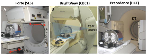
Figure 1.

The dual-head SPECT systems, the anthropomorphic phantom and the motion platform used in this project are shown. A Philips Forte: two scanning line-sources (SLS) of 153Gd scan the body along the axial direction (indicated with red arrows) obtaining transmission images simultaneous with emission imaging at every projection angle. Phantom with large breast attachment is shown. B Philips BrightView: The cone beam CT (CBCT) source, x-ray detector, and SPECT camera heads are shown in position for the 1 minute duration CT acquisition performed prior to the emission acquisition. Once the CT scan is complete, the SPECT detectors are repositioned at 90° to each other for the cardiac emission scan. C Philips Precedence: 16-slice helical CT (HCT) is used to acquire projections over the entire torso during a 6 seconds interval. For ease in viewing all of the components, the picture was taken prior to the insertion of the phantom into the CT gantry for imaging. After HCT the SPECT camera heads are also repositioned at 90° to each other for the cardiac emission scan.
