Figure 2.

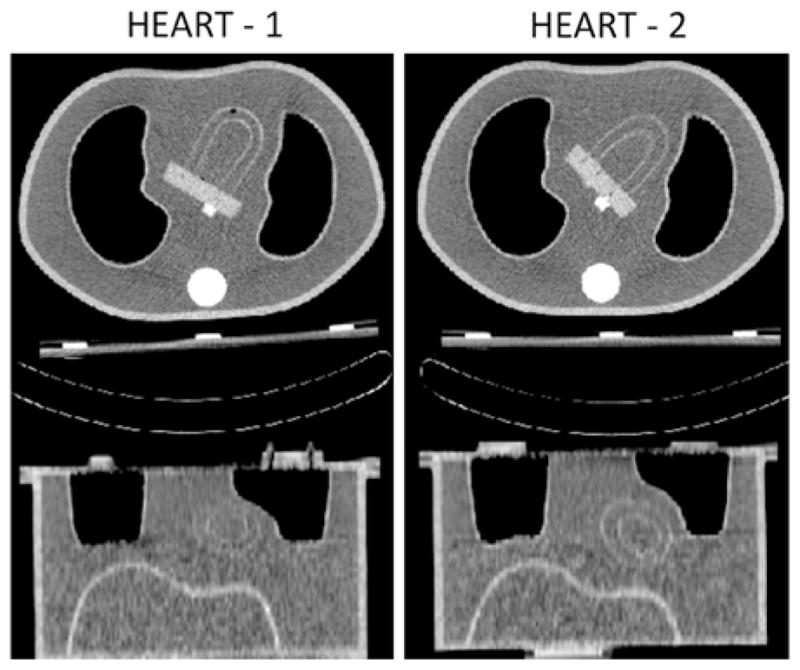
The two heart orientations employed herein are shown with transaxial (top) and coronal (bottom) slices obtained from HCT acquisitions of the stationary phantoms. A small air bubble can be seen close to the apex of the Heart-1 (left).

The two heart orientations employed herein are shown with transaxial (top) and coronal (bottom) slices obtained from HCT acquisitions of the stationary phantoms. A small air bubble can be seen close to the apex of the Heart-1 (left).