
Fig. 2. Cell death detection by PI staining and the kinetics of ZnO dissolution in solution.
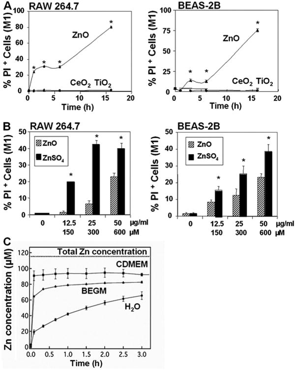
(A) After exposure to NPs suspended in tissue culture medium at 25 μg/ml for 1-16 hrs, cells were stained with 47.5 μg/ml PI and immediately analyzed in a LSR flow cytometer. The % PI-positive (M1-gated) cells were scored by Cellquest. (B) PI uptake in response to exposure to 12.5-50 μg/ml ZnO and an equimolar concentration of Zn in the form of a ZnSO4 solution for 6 hr. The maximum molar concentration of Zn added to these cultures in particulate or suspended form were 150, 300, and 600 μM, respectively. (C) The kinetics of ZnO nanoparticle dissolution in water and the two cell culture media: (i) complete DMEM containing 10% FBS and (ii) BEGM containing growth factors, cytokines, and supplements. Each data point is the average of three replicates, with error bars denoting the standard deviation.